

Pterygium
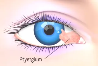
A pterygium is a triangular-shaped lesion that grows from the conjunctiva (the thin tissue on the white surface of the eye) towards the cornea. This lesion is caused by a benign growth of connective tissue and blood vessels in the conjunctiva.
In the early stages, the pterygium appears as a white area and gradually progresses towards the center of the cornea. Due to the presence of many blood vessels, its color is usually pink or red.
Symptoms of pterygium
- Burning and itching
- Red and watery eyes
- Reduced vision in advanced cases
- Causes astigmatism even in small pterygiums
In some cases, the pterygium becomes so large that it advances to the middle of the cornea and blocks vision.
Factors contributing to the development of pterygium
Ultraviolet (UV) radiation: prolonged exposure to the sun
Strong wind and dust: chronic eye irritation
Prevention: Wearing sunglasses and a brimmed hat is recommended for people who work or live in bright and dusty environments.
Treatment of pterygium
The main treatment for pterygium is surgery. However, in some cases, there is a possibility of its recurrence.
If only the lesion is removed during surgery and the surface underneath it is left uncovered, there is a possibility of recurrence in about 80% of cases. However, in new methods, the surface underneath the lesion is covered with connective tissue (graft) to reduce the possibility of recurrence.
Pterygium surgery methods:
1. Conjunctival autograft surgery (CAG)
In this method, a part of the patient’s own conjunctiva is separated from a healthy area and placed at the site of the lesion.
2. Amniotic membrane surgery (AMT)
This procedure uses amniotic membrane (the placenta of a newborn baby) that the doctor obtains from a tissue bank. This procedure is useful for covering the damaged area and reducing the chance of recurrence.