


Retina
Retinal Detachment
The retina is a thin layer of light-sensitive nerve tissue located at the back of the eyeball. This layer is responsible for converting light into nerve messages and sending them to the brain; therefore, it plays a very important role in the visual process. The retina depends on a vascular layer called the choroid to receive oxygen and nutrients. If the connection between the retina and the choroid is cut off, the retina’s function is impaired due to lack of oxygen and the person’s vision is reduced. If this condition persists, the retinal tissues undergo necrosis (tissue death), which is irreversible. For this reason, retinal detachment is considered an ophthalmological emergency.
Retinal Detachment Treatments
1. Laser Retinal Bonding or Freezing (Laser Retinopexy)
In this method, the holes in the retina are burned using a laser (from inside the eye) or cryotherapy (from outside the eye). These burns cause adhesion between the retina and the wall of the eye and prevent fluid from entering the back of the retina. This method is used only in cases where the retina is perforated but has not yet detached. This treatment is performed on an outpatient basis and with local anesthesia.
2. Pneumatic Retinal Bonding (Pneumatic Retinopexy)
In this method, after local anesthesia, a gas bubble is injected into the eye. This bubble applies pressure to the retina and adheres it to the wall of the eye. It also prevents fluid from leaking behind the retina. This method is only effective for detachments in the upper part of the retina. The patient must maintain a certain position of the head and body for a while (such as sitting or lying down). The gas bubble is absorbed within 10 to 14 days, and in the next step, the retinal hole is completely closed with laser or cryotherapy.
3. Scleral Buckle
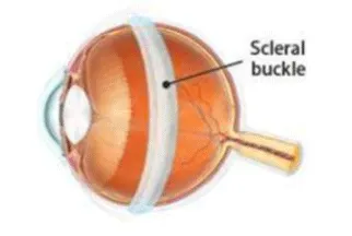
The most common method of treating retinal detachment. In this method, a strip or silicone sponge is sutured to the outside of the eye (sclera) to push the wall of the eye inward and bring the detached retina closer to its place. During the procedure, the fluid accumulated behind the retina is drained and the retinal holes are also closed with laser or cryotherapy. This buckle is permanent, not visible from the outside and does not change the appearance of the eye, but it may cause nearsightedness.
4. Vitrectomy
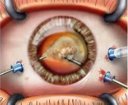
This surgery is performed to treat retinal and vitreous disorders such as diabetic retinopathy, macular hole, severe eye injuries, and some cases of retinal detachment. In this procedure, the vitreous (the jelly-like substance inside the eye) is drained and replaced with a special gas or liquid to reposition the retina.